Biology Forum › Cell Biology › Nuclei With Holes in Them
- AuthorPosts
- April 17, 2010 at 7:30 am #13125
 TecmoParticipant
Hey guys,
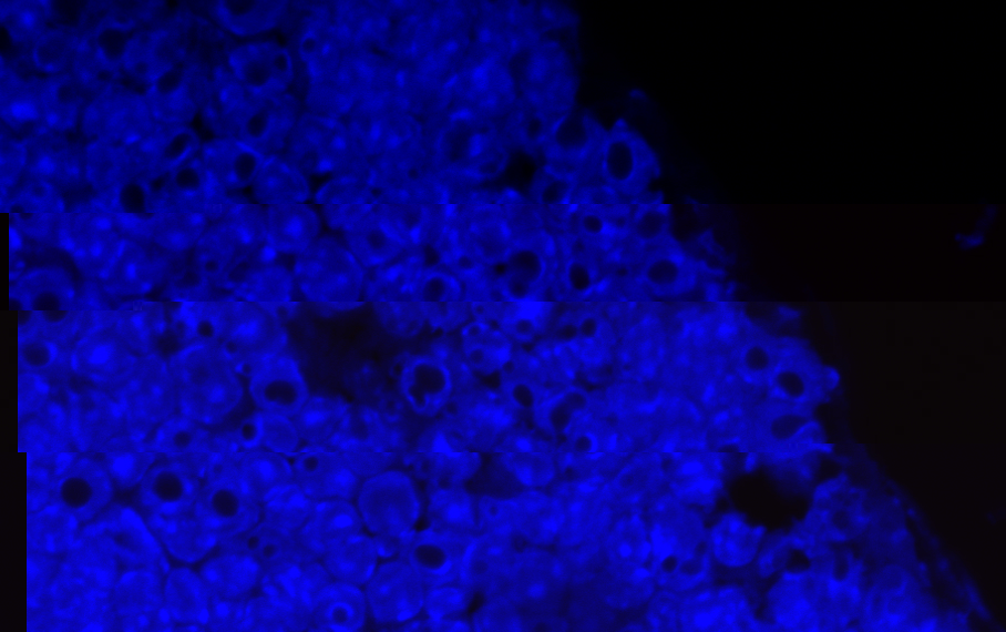
I got this imaging DAPI stained cells the subcapsular zone of an adult mouse thymus. Note the cells with hole sin the nucleus. I currently have no idea what it means. A quick google search didn’t bring up anything useful either. Does anyone have any insight they could provide?
Thank you for your time.
Attachments:

- April 17, 2010 at 7:57 am #99072JackBeanParticipant
To me it seem like if you had stained everything except nucleus.
The correct DAPI staining should not result with everything blue, but not discrete nuclei.
http://www.servlab.co.kr/exalpha/T105M.jpg
Notice the second pictures from top. THe green is whole cell, whereas blue is only nucleus.
The small dots in nuclei in third line are IMHO nucleoli (but can there be two or more per nucleus?) - April 17, 2010 at 8:08 am #99075TecmoParticipant
Hey Jack,
I don’t think it’s staining the whole cell, it’s just there is a high cell density in the thymus and developing thymocytes have huge cell nuclei. You might be on to something with the nucleoli, perhaps it is enlarged for some reason?
- April 17, 2010 at 8:42 am #99076JackBeanParticipant
I have seen now a picture in Albert’s The Cell and there are some proteins (fibrinogens?), which are located in the the nucleolus and Cajal bodies and I think it did seem very similar.
- April 17, 2010 at 11:45 pm #99091TecmoParticipant
Enlarged Cajal bodies seem a lot more likely now. After reading a bit about them, they’re supposedly enlarged in cells with high rates of transcription. These cells in the sub-capsular zone are developing thymocytes. Although, many thymus cells are developing thymocytes so I have no idea why this is only happening here. The only thing I can think of is that the DN4 to ISP transition may put mor eload on the cell transcription wise, in comparison to DN1-2-3 transitions or ISP to DP.
- AuthorPosts
You must be logged in to reply to this topic.
No related posts.