Biology Forum › Microbiology › Microscopy questions
- AuthorPosts
- January 29, 2006 at 7:49 pm #3398
 LuciParticipant
Hi,
I am enrolled in a Biology class as my uni made it mandatory for us to complete a science course. I figure I could do well in biology as compare to other science. So far, I am motivated to learn, but have some difficulties. Perhaps some of the experts out there are able to help me.
Could you please explain to me the concept of "depth of field?"
Related to this concept, i am supposed to answer the following question:1. Explain why only one of the threads, on the slide with 3 colored threads, was in focus at any given time under high power magnification, but all 3 threads appeared to be in focus at low power?
My answer is: At low power magnification the field of view is larger which allows for all 3 threads to be in view. As the power magnification increases, the field of view decreases and we can only see only one thread.
Is my answer accurate?
2. Consider the elodea cells you viwed in lab. What two structures found in these cells would distinguish it as a plant cell when comparing it to the cheek cells you viewed.
My answer is: The two structures are the regular cell wall and the chloroplast. The animal cell does not have a wall, is irregular, and does not have chloroplast.
But, again I’m not sure if this is the right answer.3. There are a couple of basic ways to increase the contrast of an image to make the specimen easier to view in the microscope. One is to reduce the amount of light with the iris diaphragm. Name another way.
I’ll say by using the rheostat (reduce the amount of light with the rheostat?) or by using stains.
It will be very helpful if you could help me with these questions.
Thank you,
Luci - January 30, 2006 at 8:49 am #38892Dr.SteinParticipant
No.1 – 🙄
No.2 – Yep, correct
No.3 – 🙄
Aww shame on Dr.Stein 😳 😛
- January 30, 2006 at 10:45 pm #38972LuciParticipantquote Dr.Stein:No.1 – 🙄
No.2 – Yep, correct
No.3 – 🙄
Aww shame on Dr.Stein 😳 😛
Dr. Stein,
The rolling eyes for 1 and 3. Does that mean I’m wrong? And what would make my answer better?
Thanks a million,
Luci - January 31, 2006 at 6:42 am #39032Dr.SteinParticipant
Nah, those rolling eyes means that I do not know the answer, that’s why my face turns into red 😀 😳 <— like this
- February 3, 2006 at 1:57 am #39397Ken RamosParticipant
Question No. 1 You are basiclly right. However the numerical aperature (n.a.) of the microscope objective also comes into play here. N.A. is the most important indicator for the quality of a microscope objective. It determines the speed of the lens and also relates to the resolving power. At low magnification you are right, field of view increases and so does the objectives n.a., the ability to resolve all three threads at once. As the magnification increases, you are right again, the field of view decreases and so does the objectives ability to resolve all three threads but one. So, your answer is good but remember the numerical aperature or N.A./n.a. plays an important roll also. 😉
Question No. 2 Dr. Stein is right. 😀
Question No. 3 There are many ways to increase and decrease contrast in a microscope. The condenser iris is but one of them. Clear blue filters or various other colored filteres, placed in the filter tray below the condenser is another, your rheostat is one, focusing your condenser is another. Then there is Kohler illumination, which my microscope has, plus all the above, in adjusting contrast also. Your in the ballpark with your answer but it is vauge. 🙄 Those items listed above should be of help to you. 😀
Some others of note but no need to dwell on them at this time, is Phase Contrast, Darkfield imaging and Circular Oblique illumination (COL). DIC or Differential Interfernce Contrasting is another. A new technique recently introduced by Zeiss of Germany is VAREL Contrasting or Variable Relief Contrasting. This has the combined features of COL and Phase Contrast. 😮
Ask your professor or teacher to further exlain these techniques to you. You may find them extremely interesting. 😀
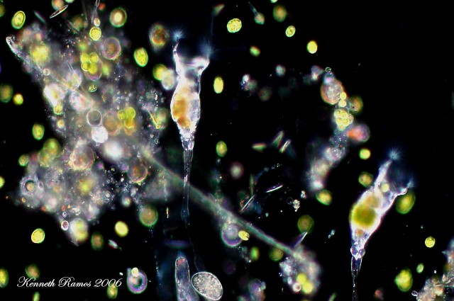
Here is an example of Darkfield Imaging and contrasting. The subject a Rotifer(s), species Collotheca. 😀
Attachments:

- September 16, 2010 at 3:25 am #101290scriptParticipant
Hi,
I would like to know more regarding depth of field. For example you have 3 threads, A, B and C. A is at the bottom, B is on top of A and C is on top of B. During 4x magnification, all thread can be seen and focus. During 10x magnification, only thread B and C are focus. During 40x magnifaction, only thread C is focus.
I have a doubt. During 10x magnification, will there be an event whereby only thread B is focus and thread C and A are not focus. Similarity will there be an event whereby only thread C is focus and thread B and A is not focus.
During 40x magnification can other thread be focus instead of thread C?
During higher magnification, can the bottom thread to be more focus instead?
- September 20, 2010 at 6:22 am #101342JackBeanParticipant
it depends just on you, what you focus on
- AuthorPosts
You must be logged in to reply to this topic.